
PDB Structures

87. MsmUdgX H109S/Q53A double mutant. PDB ID: 8IIR
|

86. MsmUdgX H109S/R184A double mutant. PDB ID: 8IIS
|
85. Complex form of MsmUdgX H109S/R184A double mutant and uracil- obtained from uracil DNA (ttUtt) post its cleavage by MsmUdgX H109S/R184A. PDB ID: 8IIT
|
84. H109Q mutant of uracil DNA glycosylase X. PDB ID: 8IIO
|
83. Complex form of MsmUdgX H109Q mutant and uracil- obtained from uracil DNA (ttUtt) post its cleavage by MsmUdgX H109Q. PDB ID: 8IIP
|
82. MsmUdgX H109S/E52N double mutant. PDB ID: 8IIQ
|
81. Complex form of MsmUdgX H109G mutant and uracil- obtained from uracil DNA (ttUtt) post its cleavage by MsmUdgX H109G. PDB ID: 8IIL
|
80. H109K mutant of uracil DNA glycosylase X. PDB ID: 8IIM
|
79. Complex form of MsmUdgX H109K mutant and uracil- obtained from uracil DNA (ttUtt) post its cleavage by MsmUdgX H109K. PDB ID: 8IIN
|
78. H109C mutant of uracil DNA glycosylase X. PDB ID: 8IIH
|
77. Complex form of MsmUdgX H109C mutant and uracil- obtained from uracil DNA (ttUtt) post its cleavage by MsmUdgX H109C. PDB ID: 8III
|
76. H109G mutant of uracil DNA glycosylase X. PDB ID: 8IIJ
|
75. Complex form of MsmUdgX and uracil- obtained from uracil DNA (ttUtt) post its cleavage by MsmUdgX. PDB ID: 8IIE
|
74. H109A mutant of uracil DNA glycosylase X. PDB ID: 8IIF
|
73. Complex form of MsmUdgX H109A mutant and uracil- obtained from uracil DNA (ttUtt) post its cleavage by UdgX H109A. PDB ID: 8IIG
|
72. Structure of the M. tuberculosis HtrA N269A mutant at room-temperature.
PDB ID: 7W4R |

71.Structure of the M. tuberculosis HtrA S363A mutant at room-temperature.
PDB ID: 7W4S |
70. Structure of the M. tuberculosis HtrA S367A mutant at room-temperature.
PDB ID: 7W4T |

69. Structure of the M. tuberculosis HtrA S407A mutant at room-temperature.
PDB ID: 7W4U |

68. Structure of the M. tuberculosis HtrA S413A mutant at room temperature.
PDB ID: 7W4V |

67. Structure of the M. tuberculosis HtrA K436A mutant at room-temperature.
PDB ID: 7W4W |

66. Structure of the M. tuberculosis HtrA S367A mutant.
PDB ID: 7VYZ |

65. Structure of the M. tuberculosis HtrA S407A mutant.
PDB ID: 7VZ0 |
64. Structure of the M. tuberculosis HtrA N269A mutant.
PDB ID: 7W21 |

63. Structure of the M. tuberculosis HtrA K436A mutant.
PDB ID: 7W22 |
62. Structure of the M. tuberculosis HtrA S363A mutant
PDB ID: 7W23 |

61. Structure of the M. tuberculosis HtrA N383A mutant.
PDB ID: 7W24 |

60.Structure of the M. tuberculosis HtrA S413A mutant.
PDB ID:7W25 
57. Structure-guided studies of the Holliday junction resolvase RuvX provide novel insights into ATP-stimulated cleavage of branched DNA and RNA substrates. PDB ID: 7ESS
54. Crystal structure of Mycobacterium tuberculosis HtrA1 (Rv1223) in regulated conformation.
PDB ID: 6IEO |
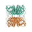
59. Crystal structure of the single-stranded dna-binding protein from Mycobacterium tuberculosis- Form III. PDB ID: 7F5Y
56. Structure of RNase J1 from Staphylococcus epidermidis.
PDB ID: 6K6S 
53. Crystal structure of PNPase from Staphylococcus epidermidis.
PDB ID: 5YJJ |

58. Crystal structure of the single-stranded dna-binding protein from Mycobacterium tuberculosis- Form III. PDB ID: 7F5Z
55. Structure of RNase J2 from Staphylococcus epidermidis.
PDB ID: 6K6W 
52. Crystal structure of Mycobacterium tuberculosis extracytoplasmic function sigma factor SigJ.
PDB ID: 5XE7 |
51. Crystal structure of Bacillus subtilis BacC Dihydroanticapsin 7-dehydrogenase in complex with NADH. PDB ID:5ITV
|
50. Crystal structure of Bacillus subtilis BacC Dihydroanticapsin 7-dehydrogenase.
PDB ID: 5ITW |
49. Crystal structure of Dihydrodipicolinate Synthase from Mycobacterium tuberculosis in complex with alpha-ketopimelic acid. PDB ID: 5J5D
|

48. Crystal structure of AgrA LytTR domain in complex with promoters.
PDB ID: 4XQQ |

47. Crystal structure of AgrA LytTR domain in complex with promoters.
PDB ID: 4XQJ |
46. Crystal structure of AgrA LytTR domain in complex with promoters.
PDB ID: 4XQN |
45. Structure of AgrA LytTR domain in complex with promoters.
PDB ID: 4XXE |
44. Structure of AgrA LytTR domain
PDB ID: 4XYO |

43. Structure of AgrA LytTR domain in complex with promoters
PDB ID: 4XYQ |
42.Crystal structure of S. aureus Homoserine Dehydrogenase at pH7.0. PDB ID: 4PG6
|
41. Crystal structure of S. aureus Homoserine Dehydrogenase at pH6.0. PDB ID: 4PG4
|
40. Crystal structure of S. aureus Homoserine Dehydrogenase at pH6.5. PDB ID: 4PG5
|
39. Crystal structure of S. aureus Homoserine Dehydrogenase at pH7.5.
PDB ID: 4PG7 |
38. Crystal structure of S. aureus Homoserine Dehydrogenase at pH8.5.
PDB ID: 4PG8 |
37. Crystal structure of ATP binding domain of AgrC from Staphylococcus aureus.
PDB ID: 4BXI |
36. Structure of Mycobacterium tuberculosis extracytoplasmic function sigma factor SigK in complex with the cytosolic domain of its cognate anti-sigma factor RskA. PDB ID: 4NQW
|
35. Crystal structure of YwfH, NADPH dependent reductase involved in Bacilysin biosynthesis.
PDB ID: 3U49 |
34. Crystal structure of YwfH, NADPH dependent reductase involved in Bacilysin biosynthesis.
PDB ID: 3U4C |
33. Crystal structure of YwfH, NADPH dependent reductase involved in Bacilysin biosynthesis. PDB ID: 3U4D
|
32. The crystal structure of a putative aminohydrolase from methicillin resistant Staphylococcus aureus. PDB ID: 4EWT
|
31. Crystal structure of SigD4 in complex with its negative regulator RsdA. PDB ID: 3VEP
|
30. Crystal structure of -35 promoter binding domain of SigD of Mycobacterium tuberculosis. PDB ID: 3VFZ
|
29. Crystal structure of the catalytic domain of PTP10D from Drosophila melanogaster. PDB ID: 3S3E
|
28. Crystal Structure of the catalytic domain of PTP10D from Drosophila melanogaster with a small molecule inhibitor Vanadate. PDB ID: 3S3F
|
27.Crystal structure of the catalytic domain of PTP10D from Drosophila melanogaster with a phosphopeptide substrate GP4. PDB ID:3S3H
|
26. Crystal structure of the catalytic domain of PTP10D from Drosophila melanogaster with a small molecular inhibitor para-NitroCatechol Sulphate. PDB ID: 3S3K
|
25. The Crystal Structure of Dihydrodipicolinate reductase from Staphylococcus aureus. PDB ID: 3QY9
|
24. Crystal structure of Staphylococcus aureus nucleoside diphosphate kinase
PDB ID:3Q83 |
23. Crystal structure of Staphylococcus aureus nucleoside diphosphate kinase complexed with GTP. PDB ID: 3Q86
|
22. Crystal structure of Staphylococcus aureus nucleoside diphosphate kinase complexed with CDP. PDB ID: 3Q89
|
21. Crystal structure of Staphylococcus aureus nucleoside diphosphate kinase complexed with ADP and Vanadate.
PDB ID: 3Q8Y |
20. Crystal structure of Staphylococcus aureus nucleoside diphosphate kinase complexed with ADP. PDB ID: 3Q8U
|
19. Crystal structure of Staphylococcus aureus nucleoside diphosphate kinase complexed with UDP. PDB ID: 3Q8V
|
18. Crystal structure of Staphylococcus aureus metallopeptidase (Sapep/DapE) in the apo-form. PDB ID: 3KHX
|
17. Crystal Structure of R350A mutant of Staphylococcus aureus metallopeptidase (Sapep/DapE) in the apo-form. PDB ID: 3KHZ
|
16. Crystal structure of Staphylococcus aureus metallopeptidase (Sapep/DapE) in the Mn2+ bound form. PDB ID: 3KI9
|
15. Crystal Structure of the PAS domain of Rv1364c. PDB ID: 3KX0
|
14. Crystal structure of BacB, an enzyme involved in Bacilysin synthesis, in triclinic form. PDB ID: 3H9A
|
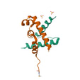
13. Crystal structure of Mycobacterium tuberculosis anti-sigma factor RslA in complex with -35 promoter binding domain of sigL.
PDB ID: 3HUG |
12. Structural and substrate-binding studies of pantothenate synthenate (PS)provide insights into homotropic inhibition by pantoate in PS's. PDB ID: 3GUZ
|
11. Crystal structure of Rv2704, a member of highly conserved YjgF/YER057c/UK114 family, from Mycobacterium tuberculosis. PDB ID: 3I7T
|
10. Crystal structure of Penicillin binding protein 4 from Staphylococcus aureus COL in complex with Cefotaxime. PDB ID:3HUM
|
9. Crystal structure of Penicillin binding protein 4 from Staphylococcus aureus COL in complex with Ampicillin. PDB ID: 3HUN
|
8. Crystal structure of BacB, an enzyme involved in Bacilysin synthesis, in monoclinic form. PDB ID: 3H7J
|
7. Crystal structure of BacB, an enzyme involved in Bacilysin synthesis, in tetragonal form. PDB ID: 3H7Y
|
6. Structure of the catalytic domain of the chick retinal neurite inhibitor-Receptor Protein Tyrosine Phosphatase CRYP-2/cPTPRO
PDB ID: 2PI7 |
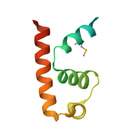
5. Crystal structure of the Pribnow Box recognition region of SigC from Mycobacterium tuberculosis.
PDB ID: 2O7G |
4. Crystal structure of the "-35 element" promoter recognition domain of Mycobacterium tuberculosis SigC
PDB ID: 2O8X |
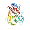
3. Crystal structure of YxaG, a dioxygenase from Bacillus subtilis
PDB ID: 1Y3T |
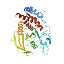
2. Crystal Structure of Mycobacterium tuberculosis NusA
PDB ID: 1K0R |

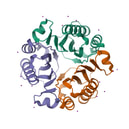
1. Crystal structure of NusB from Mycobacterium tuberculosis
PDB ID: 1EYV |
